Tuesday, December 23, 2008

Young woman with pregnancy-associated dermatosis

Abstract: 31 yo woman with atypical blood vessels on right arm and chest.
History: This healthy 31 yo woman has had an asymptomatic slowly progressive erythematous process on her right arm and shoulder. She is in her second trimester of pregnancy. The reddish areas are slowly getting more pronounced.
O/E: Mat-like telangiectasias on right arm and shoulder: Dermatomal distribution.
Clinical Photo(s):


Lab: N/A
Histopathology: N/A
Diagnosis: Unilateral Nevoid Telangiectasia
Questions:
Reason(s) Presented: For Interest. This is a benign entity, uncommonly encountered, but easy to recognize. The patient appreciates reassurance.
Reference: Unilateral nevoid telangiectasia (UNT) is a cutaneous condition consisting of congenital or acquired patches of superficial telangiectases in a unilateral linear distribution. Described in 1899 by Blaschko, its segmental pattern suggests a mechanism of somatic mosaicism apparent early in life or unmasked in states of relative estrogen excess, such as that in pregnancy or in chronic liver disease. Full Article

Sunday, December 14, 2008

Retroauricular Dermatitis

Abstract: 16 yo boy with 3-4 year history of retroauricular dermatitis
History: This 16-year-old boy was seen for evaluation of a retroauricular dermatitis that has been present for 3-4 years. He is in his usual state of health. He does not have a history of atopy. He does not wear glasses.
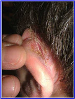
O/E: Honey-colored crusting in the superior retroauricular sulci bilaterally.
Clinical Photo:

click image to enlarge
Lab: Culture positive for many Staph. aureus with usual sensitivities.
Histopathology: N/A
Diagnosis or DDx: Retroauricular Dermatitis: This is felt to be a marker for atopic dermatitis or atopy. However, this boy is not atopic and the finding may not be all that specific. Only one article has appeared on this subject (see Reference).
Questions: Does anyone have any comments on this entity?
Reason(s) Presented: For interest.
Treatment: The patient was given a sample tube of retapamulin ointment to use b.i.d. for one week. The next photo shows appearance after one week of use as monotherapy. I plan to now use fluocinalone 0.025% ointment daily for a week or two for the residual dermatitis. This may well recur. The natural history of retroauricular dermatitis is poorly defined. There is only one article in the medical literature that discusses this entity.

status post 0ne week of retapamulin ointment

References:
Marks MB, et. al. An unsuspected sign of cutaneous allergy. J Am Acad Dermatol. 1981 May;4(5):519-22.
An eczematous eruption in the superior retroauricular areas of the scalp and often
on the posterior aspects of the pinnas may be seen in about 30% of allergic
children. The eruption is not generally noticed because the overhanging hair covers
the affected areas. The dermatitis is seen mainly in those children afflicted with
bronchial asthma, perennial allergic rhinitis, or both. A previous history of atopic
or seborrheic dermatitis is, as a rule, not elicited.

Sunday, December 7, 2008

Tuberous sclerosis


This 15-year old boy presented to my office with multiple asymptomatic papules on the face since age of three.

click on images to enlarge
Note the firm papulonodular lesions with a friable vascular surface distributed on the nose and right nasolabial fold. These lesions, called adenoma sebaceum, are actually angiofibromas - a form of cutaneous hamartoma. The patient has no neurological deficit, but tuberous sclerosis can affect many organ systems: heart, brain, kidney, liver and eyes. The lesions can be removed by light cautery and curettage or carbon dioxide laser therapy. The latter is more expensive and not much better.

A good review can be found on emedicine.com

Submitted by Henry Foong, Ipoh, Malaysia. Contact Dr. Foong.

Friday, November 28, 2008

SSSS

This is an interesting case from Ipoh, Malaysia. A large group practice like FMI will see a child with Staphyloccal Scalded Skin Syndrome occasionally. Dermatologist Henry Foong sent the pictures and the history.

HPI: This 11 mo old infant presented with symmetrical and bilateral dry scaly eruptions around the eyes and mouth for 3 days. Apparently it started with periorbital eruptions after taking some chocolates. The mother had thought it was allergy to chocolates. He has seen at least one pediatrician and a general practitioner before consulting us. There was a history of asthma. 3 other siblings had impetigo recently.

On examination, he appeared fretful. He had a low grade fever temp 37.4 degC. Marked diffuse scaling and erythema on both perioral and periorbital areas were noted bilaterally and symmetrically. The neck and axilla were similarly affected. Diffuse erythema and flushing was noted on the trunk.

Clinical Photos:

Note: The child appears irritable and uncomfortable with exudations and crusting around the mouth and eyes. Skin globally is erythematous. Healing with appropriate antibiotics is complete within 14 days.
Diagnosis: Clinically he has staphylococcal scalded skin syndrome

Lab: TWBC 12,000 N50 L36 E1 M11
Culture was negative.

Course: He was admitted to the ward and treated with IV fluids, IV cefuroxime 150mg tid and topical moisturisers. He recovered well and discharged 3 days later. Plan to do an ASOT on follow up as culture was unhelpful.

Reason(s) Presented: For interest. This is a classic case; not sure why culture was negative except that patient may have had antibiotics before it was taken.

Reference: Patel GK. Treatment of staphylococcal scalded skin syndrome. Expert Rev Anti Infect Ther. 2004 Aug;2(4):575-87.
Humans are a natural reservoir for Staphylococcal aureus. Colonization begins soon after birth and predisposes to infection. S. aureus is one of the most common causes of skin infection, giving rise to folliculitis, furunculosis, carbuncles, ecthyma, impetigo, cellulitis and abscesses. In addition, S. aureus may cause a number of toxin-mediated life-threatening diseases, including staphylococcal scalded skin syndrome (SSSS). Epidermolytic toxins released by certain S. aureus strains cause SSSS by cleaving the epidermal cell adhesion molecule, desmogelin-1, resulting in superficial skin erosion. Recent experiments have revealed similarities in the pathophysiology of SSSS and pemphigus foliaceus, an autoimmune disorder that is characterized by antibodies targeting the same epidermal attachment protein. SSSS typically affects neonates and infants but may also occur in predisposed adults. It is painful and distressing for the patient and parents, although most cases respond to antibiotic treatment. Mortality is low in infants but can be as high as 67% in adults, and is dependent on the extent of skin involvement and the comorbid state. Thus, the management of adults who develop SSSS remains a major therapeutic challenge. The antibody response against the toxins neutralizes their effect and prevents recurrence or limits the effects to the area of infection, which is known as bullous impetigo.

Monday, November 24, 2008

Dennie Morgan Lines

Abstract: 4 yo boy with severe atopic dermatitis
History: 4 yo boy with severe atopic dermatitis. He's had severe atopic dermatitis since infancy and has asthma as well
O/E: The clinical picture shows classical Dennie Morgan Lines
Clinical Photo:

scroll down for more photos...
Lab: N/A
Histopathology: N/A
Diagnosis or DDx: Dennie-Morgan Lines of Atopic Dermatitis
Questions: None
Reason(s) Presented: Strictly for interest
References: Dennie-Morgan lines are symmetrical, depression folds that lie just beneath the lower eyelids of atopic dermatitis patients. These usually present at birth or shortly after, but not in all atopic dermatititis patients. Allergic shiners, such as darkening under the eyes can also be seen, but most often, there is periorbital edema/swelling and lichenification/hardening of the skin.

Uehara M. Infraorbital fold in atopic dermatitis. Arch Dermatol. 1981 Oct;117(10):627-9
An infraorbital fold (Dennie-Morgan fold) occurred in 74 (25%) of 300 patients with atopic dermatitis. Lower eyelid dermatitis was initially seen in 69 (23%) of these 300 patients. Of the 69 patients with lower eyelid dermatitis, 57 (83%) showed the infraorbital fold. However, such a fold was seen only in 17 (7%) of 231 patients who lacked lower eyelid dermatitis. An infraorbital fold also was observed in eight of 11 patients with contact dermatitis of the lower eyelid. Thus, it is likely that an infraorbital fold develops when the lower eyelid is affected with eczematous dermatoses of diverse origins. The frequent occurrence of an infraorbital fold in patients with atopic dermatitis may be caused by the fact that the eyelid is a site of predilection for this dermatitis

11/25/08: 15 yo girl presented for eyelid dermatitis which shows Dennie-Morgan lines and atopic shiner. She has a long history of eczema and asthma.

Wednesday, November 19, 2008

Florid Rosacea

Abstract: 42 yo man with 6 month hx of facial eruption
History: This 42 yo man was recently discharged from rehab for ETOH abuse. His is on a number of psychotrophic medications. He's been bothered with facial eruption for the past six months. Has used metronidazole gel without relief.
O/E: Marked erythema of forehead, nose, nasolabial folds. There is a suggestion of hyperpigmentation.
Clinical Photo(s):

Lab: N/A
Histopathology: N/A/
Diagnosis or DDx: Rosacea
Questions: How would you approach this patient?
Reason(s) Presented: For interest
References: http://www.emedicine.com/

Saturday, November 15, 2008

Eyelid Dermatitis

Abstract: 65 yo woman with eyelid dermatitis
History: This healthy 65 yo woman has a six month history of pruritic eyelids. She has been treated with clotrimazole cream and Neosporin ointment without relief.
O/E: Erythema and scaling of upper eyelids
Clinical Photo(s)

Lab: N/A
Histopathology: N/A
Diagnosis or DDx: Upper Eyelid Dermatitis
Questions: What would you put in the differential diagnosis?
Reason(s) Presented: For Interest
References: Later

Saturday, October 18, 2008

Acanthosis Nigricans

Abstract: 11 yo girl with three year history of acanthosis nigricans
History: This is a healthy 11 y.o. girl. Her mother noticed gradual darkening of skin in neck folds, axillae and groin around three years ago. The child is Chinese. Has not had her first menstrual cycle yet, although has some breast development. She is mildly overweight (not obese). Fitzpatrick Skin Type IV. No hirsuitism.
O/E: Velvety hyperpigmentation of skin folds. There are a few skin tags in axillae.
Clinical Photo:

Lab: Insulin Level 43 (normal 3 - 28) Hgb A1C (normal) , Serum Testosterone Level 75 (normal < style="font-weight: bold;">Histopathology: N/A
Diagnosis or DDx: Acanthosis Nigricans
Questions: How would you approach this patient and initiate an appropriate work-up?
For a full discussion go to: VGRD Blog

Wednesday, October 15, 2008

A Destinctive Dermatosis

A seven year-old girl presents with a one year history of a hypopigmented streak on the lower face.


What are your thoughts?

For answer go to VGRD Blog

Wednesday, September 24, 2008

The Ugly Duckling

Presented by Helge Riemann and David Elpern

Abstract:
12 yo boy referred for evaluation of nevi
History: The boy's pediatrician referred him for evaluation of nevi. There is no pertinent family history since he is adopted. His mother feels the large lesion on the back has changed over the past year.
O/E: Healthy child. Type II skin. All nevi, except one, look similar. The "outlier lesion" measures 1 cm in diameter, has variations in color and a white area.
Clinical Photo(s):


Dermoscopic Image

Lab: N/A
Histopathology: N/A
Diagnosis or DDx: Probable Dysplastic Nevus Syndrome in a 12 yo boy.
Questions: What would you do here?
Reason(s) Presented: To discuss the concept of the "ugly duckling" (outlier lesions). Lesions which stand out as not belonging with the others a patient has deserve more attention and one should have a lower index of suspicion for biopsying.

Discussion: Dr. Riemann
Dysplastic melanocytic nevi (DMN) are a heterogenuous group of nevi with atypical clinical features, histopathologic abnormalities, or both.
The clinical features of DMN include asymmetry, varying sizes frequently larger than regular melanocytic nevi, irregular or ill-defined borders, some color variegation showing two to three shades of brown, pink, skin-colored, or red, and surface irregularities like "cobblestoning" or "fried-egg" appearance. Histopathologically, DMN can show lentiginous proliferation, elongated or bridged rete ridges, nests varying in size, nest located in papillary tips, a junctional component extending beyond the dermal component, and some degree of cytologic pleomorphism.
Several studies have shown that the risk of melanoma is directly related to the number of ordinary nevi and to the number of DMN as defined by the clinical criteria above.
Although the term "dysplastic nevus syndrome" lacks a stringent definition, it is ususally used to describe an autosomal dominant condition of increased number of ordinary and DMN. Affected individuals have an increased risk to develop melanoma and therefore, require regular skin checks. As these patients, by definition, can have many clinically atypical nevi, the challenging task for the clincian is to identify any lesions suspicious for melanoma.
The "outlier" concept describes a helpful rule of thumb to look for any DMN which appears to clearly stand out as being different from the average, baseline clinical appearance of the patient's other nevi. These "outlier lesions" should be carefully evaluated for removal. Lesions suspicious for melanoma should then be removed with a 2-5 mm margin when possible
.
References:
1. Grob JJ, Bonerandi JJ.
The 'ugly duckling' sign: identification of the common characteristics of nevi in an individual as a basis for melanoma screening. Arch Dermatol. 1998 Jan;134(1):103-4


2. Dixon AJ, Hall RS. Managing skin cancer--23 golden rules. Aust Fam Physician. 2005 Aug;34(8):669-71.
From their collective experience in Australia and the USA, dermasurgeons Anthony Dixon and Scott Hall have compiled a list of "golden rules" for general practitioners to help reduce errors and problems with skin cancer management. It is anticipated that these tips will provide a brief yet informative reference when faced with skin cancer management concerns in general practice.
This valuable refrence can be viewed as FULL TEXT HERE

3. Emedicine.com has a good chapter on Dysplastic Nevi.


Wednesday, September 10, 2008

Abstract: 9 yo girl with vascular lesion of eyelid
History: A 9 yo girl has a one month history of a papule on left lower lid. It has bled on one occasion. She saw her pediatrician who referred her to dermatology.
O/E: 5 mm red papule
Clinical Photo(s)


After procedure:

Lab: N/A
Histopathology: Pending
Diagnosis or DDx: Pyogenic granuloma
Treatment: The area was anesthetized with lidocaine + epi and the lesion was snipped off with a curved iris scissors. The base was gently cauterized. Patient's father held her hand during the procedure.
Questions:
Reason(s) Presented: This is the kind of case that FPs can handle in the office. It akes a few minutes to reassure the patient, and it's important to have a parent there. Often it can take weeks to see a dermatologist or ENT or ophthalmologist. The procedure is simple. These lesions can reoccur after electrodessication, and if so may require excision. Imiquimod to the base may prevent that.
References: Pyogenic granuloma in children: treatment with topical imiquimod.
Fallah H, Fischer G, Zagarella S.
Australas J Dermatol. 2007 Nov;48(4):217-20.
We report the successful treatment of five children with facial pyogenic granuloma using topical imiquimod 5% cream. In all cases, resolution of the lesions was achieved within 2-4 weeks. Local erythema and scaling, consistent with a typical imiquimod response, was the most commonly observed side effect. No systemic complications were observed in any of the patients. There has been no recurrence of any of the lesions to date. Small mildly erythematous or hypopigmented macules remain at this stage of follow up.
[Had I read this first, this might have been a better approach!! But the combination may be easier as it was very quick]

Tuesday, July 29, 2008

Scalp Lesion in a Child

Abstract: 14 yo boy with alopecic area since birth
History: Shortly after birth the mother noted a yellowish alopecic area on the scalp. She was told it was from a scalp electrode and thought no more about it. About a year ago, the lesion became more raised and the surface pebbly (bossilated).
O/E: Five cm oval tan/pinkish alopecic plaque with a slightly bossilated surface.
Clinical Photo(s):

Lab: N/A
Histopathology:The epidermis shows papillomatous hyperplasia. In the dermis, the numbers of mature sebaceous glands are increased. Ectopic apocrine glands are often found in the deep dermis beneath sebaceous glands.

Diagnosis or DDx: Nevus Sebaceous of Jadasshon
Reason(s): Presented: You will all see these lesions. It is satisfying to give parents a good explanation and possibly a reference (#1) to read.
References:
1. Dermnet
2. eMedicine
3. Cribier B, Scrivener Y, Grosshans E.
Tumors arising in nevus sebaceus: A study of 596 cases.
J Am Acad Dermatol. 2000 Feb;42(2 Pt 1):263-8.
The rate of malignant tumors arising on NS was very low and we did not observe such cases in children, who had associated benign tumors in only 1.7% of cases. Benign neoplasms were common and most of them occurred on the scalp; this was not a bias resulting from a longer duration before surgery. Because most tumors occurred in adults older than 40 years, our study suggests that prophylactic surgery in young children is of uncertain benefit. Clinical follow-up is probably sufficient, and even those cases with clinical changes often proved to be benign tumors or warts.

4. Santibanez-Gallerani A, Marshall D, Duarte AM, Melnick SJ, Thaller S.
Should nevus sebaceus of Jadassohn in children be excised? A study of 757 cases, and literature review.
J Craniofac Surg. 2003 Sep;14(5):658-60
The incidence of basal cell carcinoma and the need for prophylactic excision in children with nevus sebaceus of Jadassohn have been a topic of controversy. The authors performed a retrospective analysis of 757 cases from 1996 to 2002 in children aged 16 years or younger. No cases of basal cell cancer were found in the nevus sebaceus group. Recent studies in children corroborate these findings and question the need for prophylactic surgical removal of the nevus sebaceus.

Take-Home Points:
1. Most sebaceous nevi will have a benign behavior. There is no crying need for excision at a young age.
2. These lesions have a characteristic appearance and can be diagnosed clinically.
3. They get thicker and more irregular at puberty as a result of hormones acting on the sebaceous glands

Two additional patients seen in Williamstown with N. sebacsous

Thursday, July 3, 2008

Bizarre Hyperpigmented Streaks

Abstract: 30 y.o. man with three week history of hyperpigmented streaks over hip

History: The patient's brother was seen for an unrelated problem and asked if I would look at a picture on his cell phone. His brother, a 30-year-old man, recently returned from the Bahamas where he developed a pruritic inflammatory skin disorder. The cell phone image was hazy, but interesting and I asked if his brother could come in that day. He did, and anamnesis revealed that he had been drinking Corona beer with lime and remembers rubbing his abdomen at that time. Twenty-four to 36 hours later, he developed blisters and erythema at the site where he rubbed his skin. He thought it was from the limes, but his friends told him this was fanciful.

O/E: The examination showed streaky erythema on the right hip. The lesions are along the lines of where he rubbed with his fingers.
Clinical Photo(s)
Brother will cell-phone


Patient demonstrating how he remembers rubbing his abdomen.


Lab: N/A
Histopathology: N/A
Diagnosis or DDx: Phytophotocontact Dermatitis due to Lime (Rutaceae)
Questions: N/A
Reason(s) Presented: Presented for interest. It would be possible for a patient to send an image to his dermatologist from the Bahamas on his cell phone and a tentative diagnosis arrived at. Knowing that limes contain furocoumarin (a potent photosensitizer) could have led to a more timely diagnosis. If treatment was initiated very early, some of the post-inflammatory hyperpigmentation might have been avoided. At that time, wet compresses and topical corticosteroids could have shortened the course of the inflammation.
Discussion: See reference. Limes, lemons and a number of other plants contain furocoumarin. To develop phytophotocontact dermatitis, an individual needs to have contact with the agent and then get sun-exposure before washing the skin. Then, 24 - 36 hours later an exaggerated sunburn develops which heals with post-inflammatory hyperpigmentaton. The pigment change is more pronounced in darker individuals and can last many months. It usually fades over time.

Reference: Hyperpigmented Macules and Streaks

Wednesday, July 2, 2008

Dermatologic Vignette

Abstract: 75 yo woman with 3 week history of a rash.
History: Painful rash in groin. Healthy woman. Was on antibiotics around a month ago. She used a topical cream given for another purpose by her Gyn. (It turns out the cream she had been using was clobetasol -- a super potent corticosteroid)
O/E: Firey erythema of crural folds, pubic area. Studded with fine superficial pustules.
Clinical Photo(s)

Lab: KOH prep from pustule loaded with budding heasts
Histopathology: N/A
Diagnosis or DDx: Candidiasis (Candidal Intertrigo: iatrogenic)
Reason(s) Presented: For interest. Rick factors for candida intertrigo include antibiotics, diabetes, topical and oral steroids, OCP, pregnancy. This patient took antibiotics and was applying a superpotent corticosteroid to the area. The latter acts as fertilizer for the yeast.
Reference: emedicine.com

Sunday, June 8, 2008

Gouty Tophi

Abstract: 55 yo man with painful nodules on hands
History: This 55 yo laborer was seen for painful lesions of hands and associated arthritis. He was diagnosed with gout two years ago when he had an episode of podagra. Allopurinol causes an exacerbation of his pain, so he stopped taking it and maintains himself on colchicine 0.5 mg q.o.d. It is clear that he rarely sees his PCP. The patient has a history of kidney stones.
O/E: 8-10 mm in diameter nodules on some fingers. Fusiform deformities of a few fingers. Toes normal.
Clinical Photo(s):


Lab:
Histopathology: Biopsy pending. On punch biopsy the plug had a chalky look which I assume is uric acid crystals. Pathology confirms clinical impression. I will get a photomicrograph and insert it here.
Diagnosis or DDx: Tophaceous Gout

Questions: How can this man be helped?
Reason(s) Presented:
For a discussion on treatment of tophaceous gout.
References:
1. eMedicine.com
2. Jelley MJ, Wortmann R.
Practical steps in the diagnosis and management of gout.
BioDrugs. 2000 Aug;14(2):99-107.
University of Oklahoma, Health Sciences Center, Tulsa Campus, Department of Internal Medicine, Tulsa, Oklahoma, USA.
One of the earliest described conditions, gout continues to plague humanity. It is characterised by the deposition of monosodium urate crystals in the joints and soft tissue. The main clinical features of gout are hyperuricaemia, acute monoarticular arthritis, tophi and chronic arthritis, along with nephrolithiasis. Gout typically occurs in middle age and more commonly in men. Asymptomatic hyperuricaemia does not require treatment. The initial attack of acute gout usually affects a single joint, often the first metatarsal phalangeal joint. Definitive diagnosis requires demonstration of urate crystals in the joint fluid. Treatment of acute gout includes nonsteroidal anti-inflammatory drugs (NSAIDs), colchicine and corticosteroids. The most important factor in success of treatment is how quickly therapy is begun after onset of symptoms. Drug treatment of hyperuricaemia includes allopurinol, sulfinpyrazone, probenecid and benzbromarone and should be used in patients with frequent gout attacks, tophi or urate nephropathy.

Tuesday, May 27, 2008

A Dermatologic Vignette

Pictured below are the hands of a 35 year-old plumber I saw yesterday. He was not aware of a dermatitis of his feet, but inspection showed mild moccasin-type Tinea pedis on both feet and subtle onychomycosis. KOH prep from the right hand was positive for septate hyaline hyphae.

Dermatologists see two foot and one hand disease regularly. Why this affects both feet and often toe nails and only one hand is a mystery.

This man had been treated with various creams for "hand dermatitis" without relief. Knowing of the entity "Two Hand and One Foot Disease" will enable you to quickly make the right diagnosis. The question of why this occurs is still unanswered.

I prescribed ultramicrosized griseofulvin (Gris-Peg) 375 mg per day. We'll see how it works. I prefer griseofulvin to terbinafine (Lamisil) because I do not trust Novartis (the company that makes the latter). They marked Lamisil too aggressively.


Right Hand


Left Hand

Wednesday, May 14, 2008

Recurrent Eruption Buttock

Abstract: 65 yo man with recurrent eruption of left buttock

Presented by DJ Elpern

History: This 65 yo engineer lives in the United Arab Emirates and was seen while visiting Massachusetts. For the past few years, he has had pruritic and painful eruptions on his left buttock. He is well and takes no medications by mouth. He was seen by practitioners in Dubai and England. On both occasions, he was treated with oral antibiotics which were not helpful. Over the past few months, he has developed tingling sensations in his left foot and pain in the left leg.

O/E: Photos were taken by patient in the UAE. This shows confluent pustules on buttock. When seen at my office, only pigment changes and mild scarring were noted.

Clinical Photo(s)


Suggestion of grouped vesicles becoming purulent


Appearance at time of office visit. Some mild atrophic scarring and color change is noted.

Lab: None
Histopathology: None

Diagnosis: Recurrent Sacral Herpes Simplex.

Comment: The buttocks and lower back are probably the third most common area for HSV recurrence. Had someone listened to the patient's history one would have heard "recurrent episodes" which suggests HSV. Then the history of clear vesicles at the outset makes the diagnosis. Strictly speaking, this is not "genital herpes." After the mouth and genitalia, the buttocks and thighs are probably the most common sites for recurrent HSV. Sciatic pain has been associated with sacral HSV (see reference) and often these patients are worked up for renal calculi or sciatica. There may be more constant pain, similar to the post herpetic neuralgia seen after herpes zoster.
It would be nice to see the patient with an acute episode so a Tzanck smear could be done. With his travel history, this may not be practical.
Rx: The acute episodes can be treated with acyclovir or a related drug. When patients have neuralgic pain as this man does, there may be some value in long term suppressive therapy with acyclovir. Dose 400 mg tid for three or four weeks.

Reason(s) Presented: This is a relatively common disorder, although it is frequently misdiagnosed as bacterial infection or "bite." The key points are recurrent eruptions associated with itching or pain. Grouped vesicles at onset which quickly become purulent. The lesions resolve in one to two weeks. Sacral herpes is commonly associated with neuralgic pain, in my experience.

References: There are few articles on this common disorder. In 1974 RB Layzer and MA Conant published what remains until today as the most important paper on this subject.
Neuralgia in recurrent herpes simplex.
Arch Neurol. 1974 Oct;31(4):233-7.
A PDF of this article is available. Thank you to Barbara Harness, Librarian and CME Co-Coordinator at Maine General Medical Center facilitated the retrieval of this article.
Basically, it reports on four patients with neuralgic pain associated with HSV infections. One of these had sciatica as the prodrome to new episodes. More needs to be written about this entity.

Sunday, May 11, 2008

25 Year History of Foot and Hand Dermatitis

Abstract: 47 yo woman with plantar dermatitis x 25 years

Presented by Natalie Karishev and David Elpern

History: This 47 yo woman has a 25 y h/o a persistent relapsing vesicular dermatitis of the L sole and to lesser extent R sole and both palms. The dermatitis is associated with mild itching, and painful fissures. KOH prep and culture on one occasion in the distant past was positive for T. rubrum. She was treated with Lamisil po for two months without any improvement. At present the rash is managed by Triamcinolone 0.025% without significant results as well. The involved area is expending

O/E: Well defined scaly patches with deep seated vesicles on the lsft palm and localized non-inflammatory scaling on localized areas of the palms.


Clinical Photo(s)


Lab: Repeat KOH prep and fungal culture are negative (April 2008)
Histopathology: NA
Diagnosis or DDx:
Tinea pedis (appears unlikely)
vs Pompholyx, Dyshidrotic Eczema) Cronic and recurrent vesicular hand/foot dermatitis
vs Psoriasis

Reason(s) Presented: For diagnostic and treatment suggestions

Comment: One of us thinks that Fran Storrs article (referenced below) is helpful for understanding this process, although she addresses a similar condition primarily of the hands. The patient described here needs patch testing (if it has not been done) and a bit more work-up. If there are frank pustules, the diagnosis of pustulosis plantaris need to be added. If diagnostic uncertainty persists, biopsy of an acute lesion may be helpful.

References: Acute and recurrent vesicular hand dermatitis not pompholyx or dyshidrosis. Storrs FJ. Arch Dermatol. 2007 Dec;143(12):1578-80.
So now do we know what pompholyx is and what causes it? I am afraid not. I join the European Dermatolo-Epidemiology Network study’s lament that we use inexact terminology in defining all kinds of hand eczema. This lack of precision in definition has rendered accurate analysis of causation and comparisons of therapeutic strategies impossible. Thorough descriptions of methods and definitions of interpretations and relevance are lacking or are simply not adhered to. In short, as the European Dermato-Epidemiology Network study suggests, we should “start again.”
Clearly, the terms pompholyx and dyshidrosis are obsolete in that really no modern investigators use them as originally defined. Some dermatologists (old ones like me) define them rigidly for use in our own patients’ care, but in studies and reviews such rigidity seldom prevails. Until a more concise label can be agreed on, I propose the use of “acute and recur- rent vesicular hand dermatitis,.” This will not include feet-only situations, but dermatologists caring for these patients know that feet can be included in this primarily hand condition. It is time for pompholyx and dyshidrosis to exit. The full article can be obtained as a PDF from Dave Elpern.

Friday, April 25, 2008

Acanthosis Nigricans

Abstract: 76 yo woman with 6 months history seen on July 25, 2007.
History: This elderly woman presented for progressive hyperpigmentation of skin folds (neck, axillae, groin, and inframammary folds). She felt she was in good general health, but noted some recent weight loss, "bloating" and scalp pruritus. She had a sister who died of pancreatic cancer.
O/E: Velvety gray-black hyperpigmentation of affected areas. In addition some she has some verrucous tumors on arms and legs.
Clinical Photo(s):


Lab: Ultrasound of abdomen showed ascites. Abdominal CT revealed "focal masses involving the omentum and peritoneal cavity most likely representing metastatic disease. Extensive ascites, hepatic custs and a left lower lobe nodule of uncertain significance. Exploratory lap found wide-spread metastatic disease, exact cell-type could not be determined.
Histopathology: N/A
Diagnosis: Malignant Acanthosis Nigricans.
Reason Presented: For interest.
Discussion:"The exact incidence of AN is unknown. In an unselected population of 1412 children, the changes of AN were present in 7.1%. Obesity is closely associated with AN, and more than one half of the adults who weigh greater than 200% of their ideal body weight have lesions consistent with AN. The malignant form of AN is far less common, and, in one study, only 2 of 12,000 patients with cancer had signs of AN.
Malignant AN is associated with significant complications because the underlying malignancy is often an aggressive tumor. Average survival time of patients with signs of malignant AN is 2 years, although cases in which patients have survived for up to 12 years have been reported. In older patients with new onset AN, most have an associated internal malignancy." (from eMedicine chapter)
Follow-up: The patient was seen back on April 18, 2008. She has had chemotherapy and is doing OK. She was again complaining about the hyperpigmentation. She looked pretty good; but understands she has metastatic disease of unknown origin. She was looking forward to going to Florida in a few weeks to visit a daughter. She would like the dark areas treated. Tretinoin may be of some value and will be tried when she returns. In the face of a grave prognosis she is a feisty septuagenarian with a smile and a sense of humor.
References: eMedicine Chapter on A.N.

Thursday, April 17, 2008

Perplexing Dermatosis of Legs

Abstract: 33 year-old man with perplexing rash on legs and feet
Presented by Natasha Karishev and Dave Elpern

HPI: This 33-year old man was seen at the FMI on April 11, 2008. He has noticed an asymptomatic eruption on legs and feet for the past few months. He had Graves Disease treated over ten years ago and now is hypothyroid. In addition, he suffers with hypertension and bipolar disorder. His medications include: HCTZ , Depakote, Levothyroxine, Imipramine, Zolpidem, Caduet ((Amlodipine/Atorvastatin)

O/E: Both lower legs are covered with orange-brown, some rust-colored, speckled, cayenne pepper–like discoloration. Spared area under socks. The “Cayenne Pepper-Like” spots are punctate areas of hemorrhage. The patient has exophthalmos, a residual effect of his past Grave’s disease.

Photos: Click on image to enlarge


Diagnosis: Pigmented Purpuric Dermatosis: Subtype: Schamberg Disease


Reason Presented: As a teaching opportunity re: an easily recognized entity that can be misdiagnosed if not known about. This is what is called “A Dermatologic Vignette.”

Discussion: From eMedicine: “The pigmented purpuric dermatoses are a group of chronic diseases of mostly unknown etiology that have a very distinctive clinical appearance. They are characterized by extravasation of erythrocytes in the skin with marked hemosiderin deposition. The etiology is unknown. Venous hypertension, exercise, and gravitational dependency are important cofactors that appear to influence disease presentation. No medical intervention is of proven benefit for the treatment of the pigmented purpuric dermatoses.
* If pruritic use topical corticosteroids and antihistamines.
* Associated venous stasis should be treated by compression stockings.
* Prolonged leg dependency should be avoided.”

In the present case, it is likely that his drugs are playing a role. These may lead to venous hypertension and resulting leakage of rbc. There are old reports of HCTZ causing PPD. The clear areas under his socks indicate that compression stockings may be the best approach here. It is thought that PPDs are caused by capillaritis which leads to leaky vessels. The extravasated RBCs are red (Cayenne Pepper Spots) and when these are reabsorbed they leave hemosiderin behing (the golden brown rusty color)

Reference: The chapter on PPD in eMedicine is an adequate introduction:

Monday, March 31, 2008

Changing Mole

Presented by: Liliana Rivis

Abstract:
14 yo boy with a changing mole.

History: 14 yo teenage boy of fair complexion, remembers having this mole for at least 5 years. He had hx of sunburn in the past 5 years during the summer. For the past year the mole started to show darker pigment in the middle, seems a little bigger, and lately was noticed to have a white halo around it.

O/E: Spongy ,elevated, dark brown with darker pigment in the middle, pretty symmetrical mole , located in the anterior mid-chest. It is surrounded by a halo. Size is 6x7 mm.

Clinical Photo(s)


Lab: N/A
Histopathology: N/A

Diagnosis or DDx: Probable Halo Nevus
Questions: Should it come out now?

References: There is a good chapter on Halo Nevus in eMedicine.com
Here is a section regarding management:

Medical Care: Halo nevi are benign, and no treatment is necessary.

Consultations: The chief diagnostic consideration in patients with halo nevi is melanoma that is undergoing regression, although making this distinction is not usually difficult. Primary melanoma is usually solitary, whereas halo nevi are commonly multiple. Furthermore, children are affected more commonly with halo nevi; adults are affected far more commonly by melanoma.

See article for more details.

Friday, March 28, 2008

10 Year History of Intertrigo

Abstract: 59 yo man with Intertrigo and AODM

History: This 59 yo disabled man has had a resistant intertrigo for ten years. He was referred by his PCP because he did not respond to miconazle and Nystatin. Meds include oral hypoglycemics, metoprolol, HCTZ, statins.

O/E: Erythematous plaques covering groin, genitalia, axillae. Complete exam revealed erythema and crusting in gluteal cleft in addition (no one had looked there).
Clinical Photo(s)

Great T-shirt!


Gluteal Cleft

Lab: KOH prep negative
Histopathology: Biopsy Pending
Diagnosis or DDx: Inverse Psoriasis
Questions/Comments: This patient was followed for a yeast intertrigo for ten years by his physicians. It was assumed to be Candida because of his diabetes. The tip off was the sharp margination and the scale. When I checked the gluteal cleft, that clinched the diagnosis. We'll see if the biopsy confirms the clinical diagnosis. In addition, it is known that beta-blockers can exacerbate psoriasis and his metoprolol may be playing a role.
Reason(s) Presented: For interest. If one thinks of the diagnosis one can do the appropriate test: a simple punch biopsy.
References:
Clinical analysis of 48 cases of inverse psoriasis: a hospital-based study.
Eur J Dermatol. 2005 May-Jun;15(3):176-8.Wang G, Li C, Gao T, Liu Y.
Department of Dermatology, Xijing Hospital, Fourth Military Medical University,
15 Changlexi Road, Xi'an 710032 China. xjwgang@fmmu.edu.cn
Inverse psoriasis, rare in clinical practice, refers to psoriasis only or mainly
occurring at flexural sites, such as the axilla, antecubital fossae, popliteal
fossae, and inguinal creases. It is also known as flexural psoriasis. With a
total collection of psoriatic cases from September 2002 to December 2003 at
Xijing hospital, we made a retrospective analysis of the disease history,
clinical characteristics, and treatment of the patients affected with inverse
psoriasis. The results showed that the major clinical manifestations of inverse
psoriasis were sharply demarcated erythematous plaques with varying degrees of
infiltration and minimal or no scales. Affected areas often involve the groin,
axilla, genitals, and umbilicus. The disease responds well to the narrow band UVB
therapy. Compared with common psoriasis, inverse psoriasis has similar and unique
characteristics in terms of the affected areas, clinical symptoms, and responses
to the treatment.